Table of Contents
- Why Peptide Degradation Matters in Research
- Visual Signs of Degraded Research Peptides
- Chemical Pathways Behind Peptide Breakdown
- How to Store Lyophilized Peptides for Long-Term Stability
- Peptide Storage Best Practices After Reconstitution
- Peptide Batch Consistency for Research Reproducibility
- Degradation vs. Inactivity: What Your Assay Results Are Telling You
- Analytical Verification: When Visual Inspection Is Not Enough
- Frequently Asked Questions
Last Updated: September 13, 2026
Why Peptide Degradation Matters in Research
A degraded peptide vial looks identical to a good one in the freezer, and the first warning is usually a failed assay three weeks into a study. That is the core problem with signs of degraded research peptides: the earliest indicators are subtle, and the expensive ones are invisible.
Peptide degradation is the loss of structural integrity through chemical or physical breakdown, reducing active compound concentration and altering assay behavior. It is not the same as a peptide being inactive by design, and conflating the two costs labs months of troubleshooting.
Visual Signs of Degraded Research Peptides
The most reliable early warning is a change from the batch’s own baseline, not a universal standard. Photograph every vial on arrival and compare against that image, not your memory.
Work through these indicators in order:
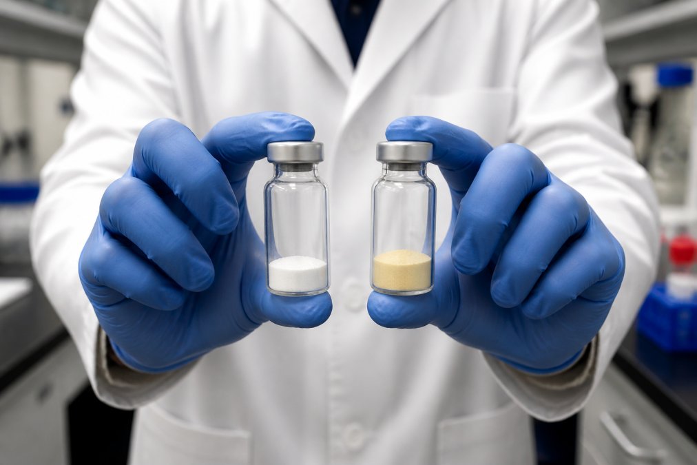
- Color shift in the powder. White to off-white is normal. Yellow, amber, or brown is not.
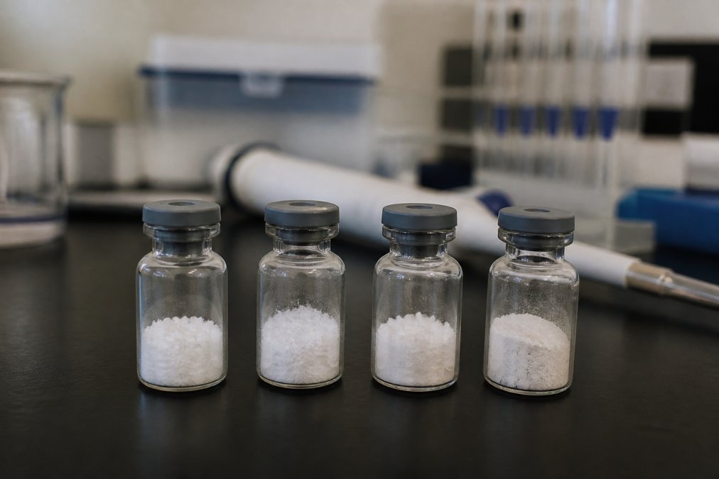
- Texture change. A healthy lyophilized cake is uniform and porous. Sticky, shrunken, or collapsed cakes indicate moisture exposure.
- Cloudiness after reconstitution. Clear solution turning hazy suggests aggregation or undissolved particulates.
- Visible particles. Any specks, fibers, or sediment in solution mean the vial is compromised.
- Incomplete dissolution. Powder that will not fully dissolve after gentle swirling has likely aggregated.
- Unusual odor. A sulfurous note often tracks with methionine oxidation.
Color Changes in Lyophilized Powder
Discoloration is the most photographed indicator and the most misread. Yellowing typically points to oxidation of Tryptophan, Tyrosine, and Methionine; browning suggests advanced breakdown, often Maillard-type reactions between residual sugars and amino groups.
A slight cream tint can be normal for certain sequences, which is why baseline photography matters more than any color chart. If your vial arrives cream and stays cream, nothing has changed; if it arrives white and turns cream in storage, something has.
Cloudiness and Particulates in Solution
Cloudiness after reconstitution is a solubility problem, and solubility problems almost always trace back to aggregation: clumped peptide molecules form larger structures that scatter light and resist dissolution.
Particulates are a harder stop. Visible particles mean the material is unsuitable for sensitive in-vitro work, because you cannot know what fraction of your intended concentration is in solution. Filtering does not fix this, it removes the evidence along with the aggregates.
Never proceed with an assay on a cloudy or particulate-containing solution “just to see what happens.” Aggregates can bind non-specifically to assay surfaces and produce results that look like real activity but are pure artifact.
Chemical Pathways Behind Peptide Breakdown
Visible changes are symptoms. The underlying chemistry runs on two main tracks, and knowing them tells you which storage variable matters for your sequence.
Oxidation and Hydrolysis
Oxidation is the modification of susceptible residues, most commonly Methionine, Tryptophan, Tyrosine, and Histidine, by reactive oxygen species including hydroxyl radicals. It is frequently metal ion-catalyzed, so trace transition metals in buffers or water accelerate it dramatically; oxygen and light both feed this pathway.
Hydrolysis is the cleavage of the peptide backbone or side-chain amide groups by water. It is the primary threat in aqueous solution and the main reason reconstituted material has a shorter stability window than lyophilized powder. Acidic or basic conditions speed it up; near-neutral pH with proper buffering slows it down.
The two pathways respond to different controls. Oxidation is managed by limiting oxygen, light, and metal exposure. Hydrolysis is managed by limiting water and controlling pH. A storage plan that only addresses one leaves the other running.
Aggregation and Its Effect on Assays
Aggregation is the physical association of peptide molecules into dimers, oligomers, and larger assemblies, driven by hydrophobic surfaces, concentration, temperature, and agitation.
The assay consequence is what matters. Aggregates reduce the effective concentration of monomeric peptide, so a vial delivers less active compound than its label implies. Worse, aggregates seed further aggregation, so a partially aggregated stock degrades faster than a fresh one.
Aggregation is also the most likely pathway to be invisible: a solution can be perfectly clear and still contain significant oligomer content. This is the gap between visual inspection and analytical verification, and where reproducibility quietly dies.
How to Store Lyophilized Peptides for Long-Term Stability
Lyophilized powder is the most stable form of any peptide and the form to buy whenever your timeline allows. Storage temperature, moisture exclusion, and light protection do the heavy lifting.
- Store at -20°C for routine work; use -80°C for sequences known to be unstable or for multi-year archives
- Keep the vial sealed with its original stopper and crimp until you are ready to use it
- Let vials equilibrate to room temperature before opening, to prevent condensation on the powder
- Protect from light; amber vials or foil wrapping both work
- Avoid repeated freeze-thaw cycles of the powder itself
Moisture is the underestimated point. Every time you open a cold vial in a warm room, water condenses onto the powder, and that water is the substrate for hydrolysis, now inside a vial you thought was dry.
Split a large lyophilized batch into single-use aliquots under dry conditions on arrival. It costs one afternoon and eliminates the repeated-open problem entirely, which is the most common cause of mid-study degradation we see.
Peptide Storage Best Practices After Reconstitution
Once a peptide is in solution, it is on a clock, shorter than most researchers assume. The biggest mistake is treating “reconstituted” as one condition. Stability depends on the diluent, sequence, concentration, container, and temperature, and those differences can change whether a study succeeds.
Why the Diluent Changes Everything
The diluent is not a neutral carrier. It sets the pH, introduces or excludes preservatives, and determines whether microbial growth is a realistic threat.
- Bacteriostatic water (0.9% benzyl alcohol): The standard choice for multi-draw vials (the FDA). The benzyl alcohol content inhibits bacterial growth, which extends the practical usable window for vials that will be entered repeatedly. It is not a sterilant and does not stop all organisms, but it meaningfully reduces contamination risk.
- Sterile water for injection: No antimicrobial protection. Appropriate only for single-use aliquots that will be consumed immediately or frozen in single-use portions.
- Acetic acid solutions (commonly 0.1%-1%): Used for peptides with poor aqueous solubility. The lower pH can improve dissolution but also shifts the hydrolysis and deamidation landscape. Some sequences are more stable here; others are less. This is sequence-specific and should be verified, not assumed.
- Buffered saline or phosphate-buffered saline: Common for assay work, but phosphate buffers can participate in degradation chemistry and are a poor choice for long-term storage of many sequences.
Post-Reconstitution Stability Windows
The table below reflects commonly reported practical windows for research-grade peptides. Treat every figure as a starting point for your own verification, not a guarantee. Sequence, concentration, and container all shift these numbers.
| Diluent | Storage Temp | Typical Practical Window | Notes |
|---|---|---|---|
| Bacteriostatic water | 2-8°C | Days to a few weeks for many sequences | Benzyl alcohol slows microbial growth; hydrolysis still proceeds |
| Bacteriostatic water | -20°C | Weeks to months | Freeze in single-use aliquots; avoid repeated freeze-thaw |
| Sterile water | 2-8°C | Hours to a few days | No preservative; use immediately or freeze in aliquots |
| Sterile water | -20°C | Weeks to months | Single-use aliquots only |
| 0.1%-1% acetic acid | 2-8°C | Sequence-dependent; often days to weeks | Verify by HPLC for your specific peptide |
| 0.1%-1% acetic acid | -20°C | Weeks to months | Acidity can slow some pathways and accelerate others |
Two rules override the table. First, use the shortest window your study can tolerate and re-verify by HPLC if the study extends past it. Second, never assume a window transfers between sequences: a peptide stable for a month in bacteriostatic water at 2-8°C may degrade in days in sterile water at the same temperature. (Source: common chemical degradation pathways for peptides)
Microbial Contamination in Multi-Draw Vials
Every entry into a vial is an opportunity for contamination, and the signs are not always obvious, some overlap with degradation:
- Cloudiness that develops after the solution was previously clear
- New particulate matter or sediment
- Unusual odor
- pH drift, if you are tracking it
- Unexplained assay variability that does not correlate with storage time
Bacteriostatic water reduces but does not eliminate this risk. Aseptic technique, wiping the stopper with an alcohol swab before each draw, and using a fresh needle every time are the practical controls. If a vial shows any contamination sign, discard it. Do not filter and reuse.
Handling and Container Practices
- Store reconstituted peptide at 2-8°C unless your protocol specifies otherwise.
- Aliquot before freezing. Never freeze and thaw a working stock repeatedly.
- Avoid vigorous vortexing. Shear and air-liquid interface exposure promote aggregation.
- Protect from light. Amber vials or foil wrapping both work.
- Use low-binding or glass containers where adsorption to plastic is a concern for low-concentration solutions.
- Track the date of reconstitution on the vial. Memory is not a record.
If your study design allows it, reconstitute only what you will use within the shortest practical window for that sequence and diluent. The cost of a fresh vial is almost always lower than the cost of a failed assay run.
When to Re-Verify
If a study extends past the practical window for your diluent and sequence, do not simply continue. Pull an aliquot and run HPLC. If purity and molecular weight still match the COA, the material is intact and you can proceed with documented justification. If purity has dropped or new peaks have appeared, the material is degraded and the data from that point forward is suspect.
Peptide Batch Consistency for Research Reproducibility
Batch-to-batch variability is the quietest threat to reproducibility, because nothing looks wrong. The powder is white, the solution is clear, and the assay still drifts.
Peptide batch consistency for research means two vials of the same catalog number, produced at different times, deliver the same active compound concentration, purity profile, and assay behavior. That requires verified analytics on every batch, not a certificate generated once and reused.
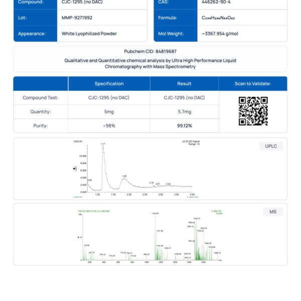
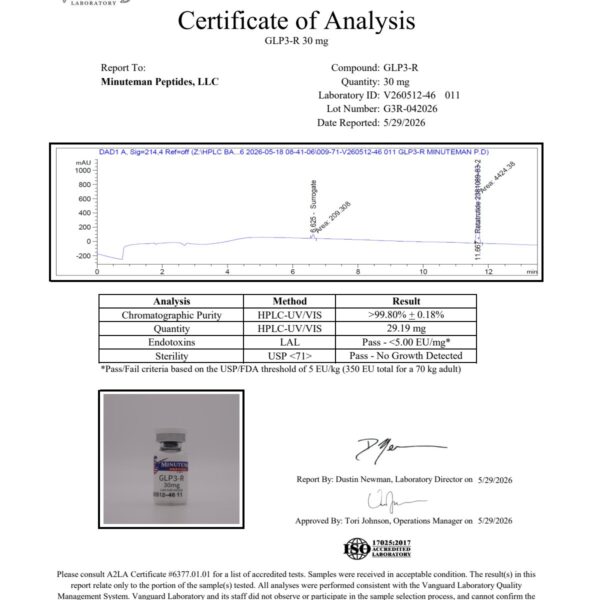
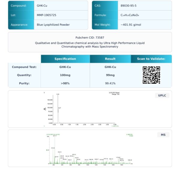
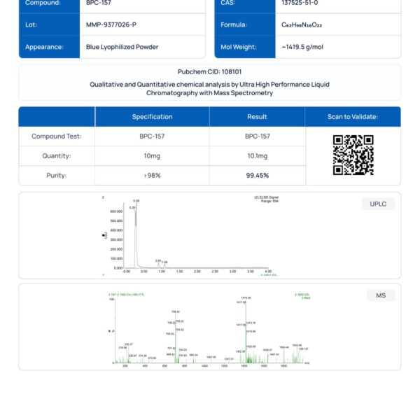
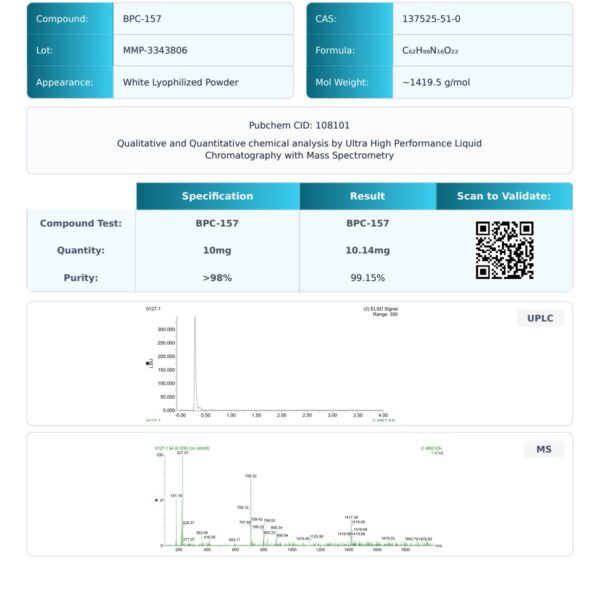
This is where documentation earns its keep. A Certificate of Analysis reporting HPLC purity and mass spectrometry confirmation from an independent ISO/IEC 17025 certified laboratory gives you a traceable basis for cross-batch comparison, and the only way to catch a subtle purity shift before it becomes unexplained variance in your data.
When planning a study that runs across multiple batches over a long period, ask your supplier how they handle batch-to-batch continuity before you commit. The answer tells you more than any single COA.
Degradation vs. Inactivity: What Your Assay Results Are Telling You
A weak assay result has at least three possible causes, and they demand different responses. Mistaking one for another sends you down the wrong troubleshooting path for weeks.
- Degradation: the peptide broke down. Purity has dropped, aggregates may be present, and the material is compromised.
- Inactivity: the peptide is intact and pure, but it does not produce the effect you expected in your specific system.
- Assay failure: the peptide is fine and active, but the assay itself is producing artifact.
The distinguishing test is analytical, not biological: run the material through HPLC and mass spectrometry. If purity and molecular weight match the COA, the peptide is intact and the problem lies in the assay or biology. If purity has dropped or new peaks appeared, you have degradation, and the question becomes what caused it.
This matters because labs routinely discard perfectly good peptide after a failed assay, then repeat the same storage mistake with the replacement.
Analytical Verification: When Visual Inspection Is Not Enough
Visual inspection catches gross degradation. It misses everything subtle, and subtle is where most reproducibility problems live. A vial can be white, dry, and clear in solution while carrying significant oxidation, deamidation, or oligomer content. The only way to know is to measure.
The Verification Stack
Different methods answer different questions. Using only one leaves blind spots.
- Reverse-phase HPLC (RP-HPLC): Separates intact peptide from degradation products and related impurities. The main peak is your target peptide; new or enlarged peaks indicate degradation products, truncations, or impurities. Purity is the main peak’s percentage of total peak area.
- Mass spectrometry (MS), typically ESI or MALDI-TOF: Confirms the main peak’s molecular weight against the expected mass. A shift of roughly +16 Da suggests oxidation; +1 Da suggests deamidation; larger shifts suggest truncation or adduct formation. MS tells you whether the main peak is actually your peptide.
- Size-exclusion chromatography (SEC) or dynamic light scattering (DLS): For aggregation concerns. SEC separates by size and quantifies oligomer content; DLS estimates particle size distribution. A clear solution can still contain significant oligomer content that RP-HPLC will not fully resolve.
- Peptide mapping or LC-MS/MS: For sequence-level confirmation of exactly where a modification occurred. This is heavier analytical work, usually reserved for critical studies.
How to Use a Third-Party Testing Service
When visual signs are absent but you suspect degradation, or you need to verify a batch before a long study, third-party testing is the answer. The workflow is straightforward:
- Select an accredited laboratory. Look for ISO/IEC 17025 accreditation, which indicates assessment against internationally recognized criteria for testing competence, the same standard referenced in a credible Certificate of Analysis (nist.gov).
- Request the right panel. For degradation verification, ask for RP-HPLC purity, mass spectrometry confirmation, and, if aggregation is a concern, SEC or DLS. Specify the expected molecular weight and sequence so the lab can compare against a reference.
- Submit a representative sample. Use a fresh, unopened vial where possible. For a suspect vial, submit it alongside a known-good reference from the same batch or a retained sample.
- Interpret the results against the COA. Compare purity, retention time, and mass to the original Certificate of Analysis. A drop in purity, retention-time shift, or mass change is evidence of degradation; a match is evidence of integrity.
- Document and act. If degraded, discard it and investigate the storage or handling cause. If intact, you have a documented basis for continuing the study.
What the Numbers Mean
A purity figure is not a pass/fail threshold on its own. A peptide at 98% purity may still contain 2% of a degradation product that is biologically active in your assay, while one at 95% may perform perfectly well if the impurities are inert. The relevant question is whether the impurity profile has changed from baseline, and whether that change matters for your assay.
This is why baseline documentation matters: a COA generated at manufacture is your reference point. Without it, you cannot know whether current purity represents degradation or simply the batch as it was made.
When to Test
- Before committing a long or expensive study to a batch
- When a vial has been in storage longer than its practical window
- When assay results drift without an obvious cause
- When a batch is received and you want to verify the supplier’s COA independently
- When you are comparing batches and need a traceable basis for the comparison
Do not rely on a supplier’s COA alone for critical work. A COA is a claim. Independent testing is verification. The two are not the same, and the gap between them is where reproducibility problems hide.
The Limits of Analytical Testing
No single method catches everything: RP-HPLC may not resolve aggregates, SEC may not resolve sequence-level modifications, and MS confirms mass but not activity. A complete picture usually requires more than one method, chosen to match the degradation pathway you are concerned about. For most research applications, RP-HPLC plus MS is the practical minimum, with SEC added when aggregation is a realistic concern.
At Minuteman Peptides, every batch is manufactured in cGMP-certified facilities and tested by an independent ISO/IEC 17025 certified laboratory, with HPLC and mass spectrometry results published transparently in the COA.
Frequently Asked Questions
How can I tell if my research peptides are degraded?
Look for color shifts in lyophilized powder (yellowing or browning), cloudiness or particles in reconstituted solution, and a gel-like texture. These visible changes often signal oxidation, hydrolysis, or aggregation. However, degradation can occur without visible signs, so analytical methods like HPLC and mass spectrometry are the only reliable way to confirm purity and potency. Always compare against a fresh reference sample when possible.
What destroys peptides during storage?
Heat, moisture, oxygen, light, and repeated freeze-thaw cycles are the main culprits. Even brief exposure to room temperature can accelerate hydrolysis and oxidation. Lyophilized peptides should stay frozen at -20°C or lower, while reconstituted peptides need refrigeration and should be used within days to weeks depending on the sequence. Proper vial sealing and desiccant use also prevent moisture intrusion.
How do I verify peptide batch consistency for research?
Request a Certificate of Analysis (COA) for each batch that includes HPLC purity data, mass spectrometry confirmation, and the testing laboratory’s ISO/IEC 17025 certification. Compare chromatograms and molecular weights across batches to ensure they match. If your lab performs its own assays, run a positive control with each new batch to confirm expected activity and rule out batch-to-batch variability.
Can I store lyophilized peptides at room temperature?
Short-term shipping at ambient temperature is usually fine, but long-term storage should be at -20°C or below. Even lyophilized peptides can absorb moisture and degrade if left at room temperature for weeks. For the best stability, keep vials sealed with a desiccant, protect them from light, and allow them to equilibrate to room temperature before opening to avoid condensation.